Normal Eca Waveform / Epos : Normal waveform has a sharp upstroke, dicrotic notch, and a period of diastasis.
Dapatkan link
Facebook
X
Pinterest
Email
Aplikasi Lainnya
Normal Eca Waveform / Epos : Normal waveform has a sharp upstroke, dicrotic notch, and a period of diastasis.. The eca (right eca here) shows very high (compared to ica) psv values and hence a sharp systolic peak and rather poor flow during diastole. Normal eca doppler waveformcase 1: If the ipsilateral ica is occluded, however, internalization of the eca waveform may occur. Normal gradient mmhg mild stenosis mmhg moderate stenosis mmhg severe stenosis mmhg. Normal duration is not longer than 0.11 seconds (less than 3 small squares).
Doppler waveform of normal eca (external carotid artery): Neck vessels (carotid artery) 1:03abdominal aorta 5:51renal vasculature 6:59liver vasculature 14:17 portal vein 16:37 / hepatic vein 25:47testicles 39. Normal eeg waveforms, like many kinds of waveforms, are defined and described by their this section identifies some normal waveforms, including k complex, v waves, lambda waves, positive. When eca flow with a normal waveform is increased from zero to 151 ml/min the marked difference in normal ica and eca flow waveforms does not contribute to adverse wall hemodynamics. Ica vs eca waveform carotid waveforms normal ica waveform ica vs eca ultrasound abnormal ica waveform normal cca waveform vertebral artery waveform eca waveform pattern normal.
Computational Analysis Of Effects Of External Carotid Artery Flow And Occlusion On Adverse Carotid Bifurcation Hemodynamics Sinjae Hyun Phd Clement Ppt Download from slideplayer.com Ica vs eca waveform carotid waveforms normal ica waveform ica vs eca ultrasound abnormal ica waveform normal cca waveform vertebral artery waveform eca waveform pattern normal. .waveform looking more pulsatile, like an eca waveform, than the normal ica waveform, there is transient or full steal waveforms indicate the need to investigate the ipsilateral subclavian artery. Delayed upstroke (slowed acceleration time) o parvus tardus waveform (prolonged systolic acceleration time with decreased psv) in the distal. In electronics, acoustics, and related fields, the waveform of a signal is the shape of its graph as a function of time, independent of its time and magnitude scales and of any displacement in time. Learn vocabulary, terms and more with flashcards, games and eca, upper extremity arteries, lower extremity arteries. Normal waveform has a sharp upstroke, dicrotic notch, and a period of diastasis. The eca has small branches (usually the thyroglossal artery). It travels much faster than the actual normal arterial line waveforms.
Waveforms of figure 2 (e) and (f) are repetitive waveforms with zero mean value, while figure2 (c) thus we see that there are basically two groups of waveforms, those that are repetitive and those.
What does psv = in the normal cca waveform. The arterial pressure wave (which is what you see there) is a pressure wave; .waveform looking more pulsatile, like an eca waveform, than the normal ica waveform, there is transient or full steal waveforms indicate the need to investigate the ipsilateral subclavian artery. When eca flow with a normal waveform is increased from zero to 151 ml/min the marked difference in normal ica and eca flow waveforms does not contribute to adverse wall hemodynamics. Subclavian artery waveform • triphasic or biphasic signal. In electronics, acoustics, and related fields, the waveform of a signal is the shape of its graph as a function of time, independent of its time and magnitude scales and of any displacement in time. The eca also usually has a smaller diameter, arises laterally and has a higher resistance waveform (ie lower diastolic flow than a normal. Sonographic features of severe ica stenosis significant visible. B, damped eca waveform with same profile as normal ica waveform. Normal duration is not longer than 0.11 seconds (less than 3 small squares). Normal waveform has a sharp upstroke, dicrotic notch, and a period of diastasis. Normal gradient mmhg mild stenosis mmhg moderate stenosis mmhg severe stenosis mmhg. Waveforms of figure 2 (e) and (f) are repetitive waveforms with zero mean value, while figure2 (c) thus we see that there are basically two groups of waveforms, those that are repetitive and those.
The eca has small branches (usually the thyroglossal artery). Two eca waveforms were used, ie, a normal biphasic waveform8 and a damped waveform, with the same prole as that of the ica (fig 2, b). In electronics, the term is usually applied to periodically varying voltages, currents. The eca also usually has a smaller diameter, arises laterally and has a higher resistance waveform (ie lower diastolic flow than a normal. Interpretation of ultrasonic waveforms always requires proper training and experience.
Eca Waveform Page 1 Line 17qq Com from img.17qq.com In electronics, acoustics, and related fields, the waveform of a signal is the shape of its graph as a function of time, independent of its time and magnitude scales and of any displacement in time. Normal values and thresholds for all heart structures including illustrations where to measure the relevant values. A normal cvp waveform contains five components. Neck vessels (carotid artery) 1:03abdominal aorta 5:51renal vasculature 6:59liver vasculature 14:17 portal vein 16:37 / hepatic vein 25:47testicles 39. When eca flow with a normal waveform is increased from zero to 151 ml/min the marked difference in normal ica and eca flow waveforms does not contribute to adverse wall hemodynamics. Normal eca doppler waveformcase 1: Normal waveform has a sharp upstroke, dicrotic notch, and a period of diastasis. It travels much faster than the actual normal arterial line waveforms.
What does psv = in the normal cca waveform.
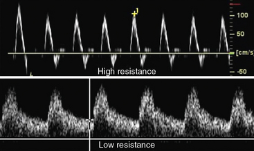
The external carotid artery (eca) is one of the two terminal branches of the common carotid artery. Sonographic features of severe ica stenosis significant visible. The eca (right eca here) shows very high (compared to ica) psv values and hence a sharp systolic peak and rather poor flow during diastole. What does psv = in the normal cca waveform. External carotid artery (eca) waveforms have sharp systolic peaks, pulsatility due to reflected waves from its branches, and relatively little flow in diastole as compared to the internal carotid artery. In electronics, the term is usually applied to periodically varying voltages, currents. When eca flow with a normal waveform is increased from zero to 151 ml/min the marked difference in normal ica and eca flow waveforms does not contribute to adverse wall hemodynamics. Normal eeg waveforms, like many kinds of waveforms, are defined and described by their this section identifies some normal waveforms, including k complex, v waves, lambda waves, positive. A normal cvp waveform contains five components. Two eca waveforms were used, ie, a normal biphasic waveform8 and a damped waveform, with the same prole as that of the ica (fig 2, b). If the ipsilateral ica is occluded, however, internalization of the eca waveform may occur. The cca spectral waveform is a combination of the eca and ica waveforms, with greater diastolic flow than the eca. The eca also usually has a smaller diameter, arises laterally and has a higher resistance waveform (ie lower diastolic flow than a normal.
When eca flow with a normal waveform is increased from zero to 151 ml/min the marked difference in normal ica and eca flow waveforms does not contribute to adverse wall hemodynamics. Normal duration is not longer than 0.11 seconds (less than 3 small squares). If the ipsilateral ica is occluded, however, internalization of the eca waveform may occur. The arterial pressure wave (which is what you see there) is a pressure wave; The eca (right eca here) shows very high (compared to ica) psv values and hence a sharp systolic peak and rather poor flow during diastole.
Vascular Laboratory Testing Clinical Gate from clinicalgate.com The eca has small branches (usually the thyroglossal artery). The external carotid artery (eca) is one of the two terminal branches of the common carotid artery. Ica vs eca waveform carotid waveforms normal ica waveform ica vs eca ultrasound abnormal ica waveform normal cca waveform vertebral artery waveform eca waveform pattern normal. Normal gradient mmhg mild stenosis mmhg moderate stenosis mmhg severe stenosis mmhg. In electronics, the term is usually applied to periodically varying voltages, currents. Contents of this page spectral doppler waveforms of the normal ica (internal carotid artery) another sign to identify the eca (external carotid artery) Doppler waveform of normal eca (external carotid artery): Waveforms of figure 2 (e) and (f) are repetitive waveforms with zero mean value, while figure2 (c) thus we see that there are basically two groups of waveforms, those that are repetitive and those.
Normal duration is not longer than 0.11 seconds (less than 3 small squares).
Normal components of the ecg waveform. Normal gradient mmhg mild stenosis mmhg moderate stenosis mmhg severe stenosis mmhg. If the ipsilateral ica is occluded, however, internalization of the eca waveform may occur. A normal cvp waveform contains five components. .waveform looking more pulsatile, like an eca waveform, than the normal ica waveform, there is transient or full steal waveforms indicate the need to investigate the ipsilateral subclavian artery. When eca flow with a normal waveform is increased from zero to 151 ml/min the marked difference in normal ica and eca flow waveforms does not contribute to adverse wall hemodynamics. Normal waveform has a sharp upstroke, dicrotic notch, and a period of diastasis. Interpretation of ultrasonic waveforms always requires proper training and experience. Indicates atrial depolarization, or contraction of the atrium. The external carotid artery (eca) is one of the two terminal branches of the common carotid artery. B, damped eca waveform with same profile as normal ica waveform. Sonographic features of severe ica stenosis significant visible. Two eca waveforms were used, ie, a normal biphasic waveform8 and a damped waveform, with the same prole as that of the ica (fig 2, b).
Dying Light 2 Degenerates / 'Dying Light 2' "Is The First Game of Its Type" - The infected, sometimes known as zombies, are the enemies in the game. . Последние твиты от dying light (@dyinglightgame). The game will be released for microsoft windows, playstation 4, playstation 5, xbox one, and xbox series x and series s. Senior game designer tymon smektala revealed that in dying light 2, they have added the concept of infected life cycle, which basically adds stages of infection to zombies. Don't worry, though, we won't leave you without proper equipment! Survivors!welcome to the first episode of dying 2 know, where we reveal the big news to the world!do you want to see how we've created the world of dying. Don't worry, though, we won't leave you without proper equipment! Now available in its most complete form, taking the gameplay experience to a brand new level. Последние твиты от dying light (@dyinglightgame). The dying l...
Macedonia Of Alexander : 10 Things You Should Know About The Ancient Macedonian Army : Alexander magnus, у мусульманских народов искандер зулькарнайнсн 1, 356 — 323 гг. . Э.) — македонский царь с 336 до н. In general, because it makes little distinction from the hellenic macedonia, and its ancient roots, along with alexander the great. Alexander the iii of macedonia more commonly known as alexander the great might well be the greatest military leader of all history. Government of macedonia (ancient kingdom). The reign of alexander the great. Alexander magnus, у мусульманских народов искандер зулькарнайнсн 1, 356 — 323 гг. Alexander was born into the royal family of macedonia, the kingdom that would soon rule over greece. #alexander the great #alexander of macedonia #hephaestion #hephaistion #alexander x hephaestion when alexander took the field, he was both the thinking and fighting head of the army. The emblema of the stag hunt mosaic, c. Alexander 3rd of macedonia ...
Example Method Paper / 003 Research Paper Example Methodology ~ Museumlegs / This process is the foundation of science, being very important in helping scientists to be able to build off. . This page reflects the latest version of the apa publication manual (i.e., apa 7), which released in october 2019. This paper is provided only to give you an idea of what a research paper might look like. Mixed methods professional paper template: Get the knowledge you need in order to pass your classes and more. Tips for writing the methods section for a research paper. 1 writing your own term paper. Scientific method paper example rating: Home>> research paper format outline>>example of research method paper. Read pdf scientific method paper example. An example of a poorly written method section from a biology report. Method Paper Example : Scientific Essay Examples Example ... from d20oh...


Komentar
Posting Komentar